
We normally use this platform to discuss the many things that can go wrong with vision. The issues you may face as you age, or if you were born a certain way are important to know and understand.
We wanted to take a different approach today and dive into how your eye’s work and what should be happening if there are no vision correction issues.
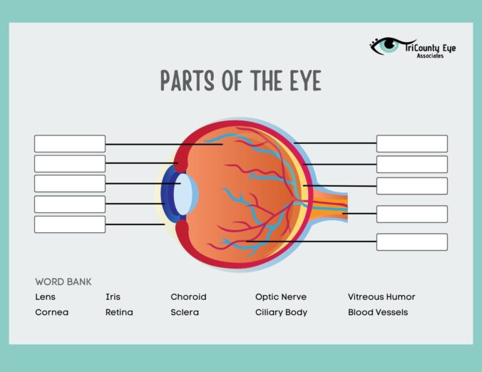
Your eye is made up of so many different parts, but they are all vital to help you see clearly. We are going to divide up the eye into four parts:
- The Eye Outside The Eyeball
- The Front of the Eye
- The Back of the Eye
- The Surface of the Eye
The Eye Outside The Eyeball
There are two main sources of connection from your eye socket to your eye ball:
- Extraocular Muscles -These are six muscles inside the eye socket (orbit) that move the eye up an down, side to side and route the eye.
- Sclera – this is a layer of strong tissue that covers almost the entire surface of the eye ball and is what the extraocular muscles attach to on the eye.
Surface of the Eye
The surface of your eye is covered with a clear membrane. This clear membrane is called the Conjunctiva, which is the mucous layer and is the first layer of tear film. The next two layers are the watery layer and the oil layer. All these layers of tears help to protect your eyes and keep them lubricated.
The Front of the Eye
The cornea is a dome shape part of your eye that light focuses into the eye through. Behind the cornea is a fluid filled space called the anterior chamber. The eye is always producing this fluid called aqueous humor. This liquid helps to regulate eye pressure by filling and draining from this space.
Behind the anterior chamber is the Iris which is the colored part of the eye as well as the pupil. The pupil is the dark hold in the middle of the eye which will constrict and dilate to control the amount of light that reaches the back of the eye.
Directly behind the pupil is the lens of the eye. The lens is what focuses the light the iris and pupil let in on the back of the eye. The cornea and lens are both really important players in making sure to give us clear vision. 70% of the eye’s focusing power comes from the cornea and 30% comes from the lens.
The Back of the Eye
There is a cavity that lies between the lens and the back of the eye which is called the vitreous cavity. It is filled with a jelly like substance called vitreous humor.
Like we already mentioned, light is focused by the cornea and lens. It is then passed through the vitreous onto the retina. The retina is the light sensitive tissue that lines the back of the eye. There is a very small area of the retina called the macula that gives us our detailed and central vision. There is also another part of the retina that provides us with peripheral vision called the peripheral retina.
The retina has cells called photoreceptors that change light into energy that is transmitted to the brain. There are two different types of photoreceptors:
- Rods – perceive black and white and enable night vision
- Cones – perceive color and provide detail vision
The retina uses electrical impulses to send light through the optic nerve to the brain. The optic nerve then sends these impulses to the visual cortex which his the part of the brain that is responsible for sight.
Conclusion
The eye is the second most complex organ in the human body, only after the brain. There are so many working parts that all work together to give you your sight. It is no mystery why it helps to have a professional help you make sure your eyes are working in tip top shape!
TriCounty Eye Associates wants to be your eye care provider and we are here for anything and everything you might need regarding your eyes!
Leave a Reply